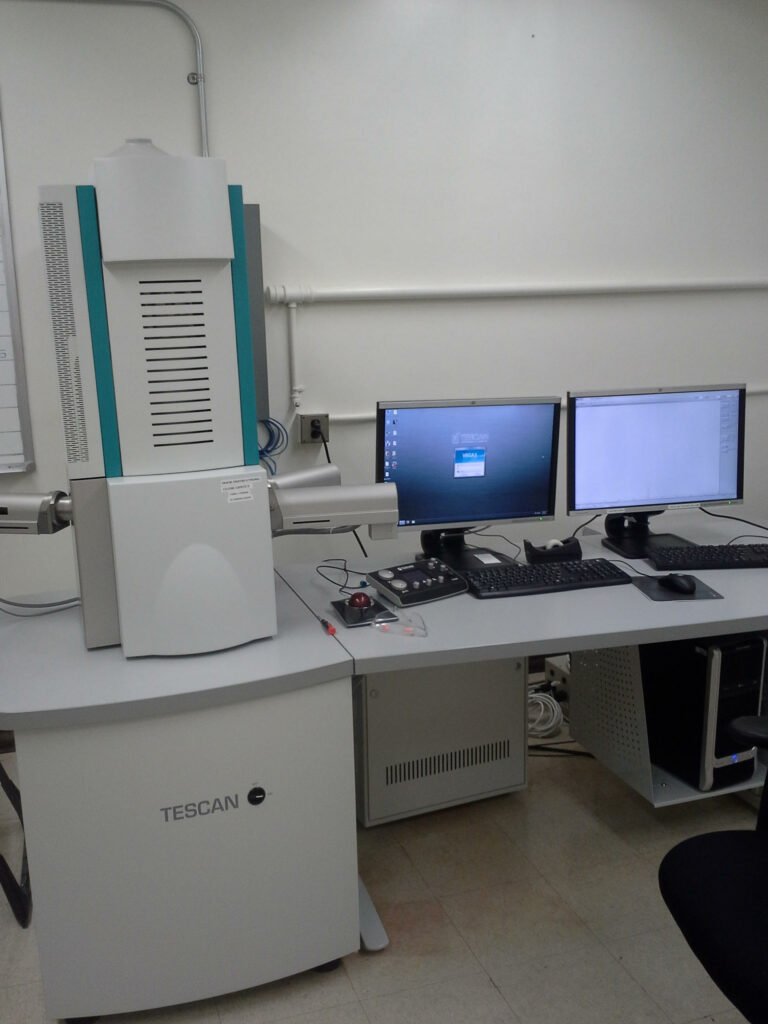
The Ion Microprobe Facility houses a Tescan Vega-3 XMU variable-pressure (VP) Scanning Electron Microscope (SEM) for the imaging and analysis of solid samples. Samples may be imaged with several detectors including a secondary electron detector (SE) for topographic imaging and backscattered electron (BSE) detector for compositional variations, along with both panchromatic and 3-channel color cathodoluminescence (CL) detectors. The SEM is also equipped with an EDAX energy-dispersive x-ray analysis (EDS) system for semi-quantitative sample compositional analysis and elemental mapping.
The sample chamber will accommodate thin sections, thick sections, ion probe mounts, pin mounts, and larger samples up to several centimeters. Samples are generally run with a conductive coating (either graphite or gold), but BSE, EDS, and CL are also available for uncoated samples in low-vacuum mode.
